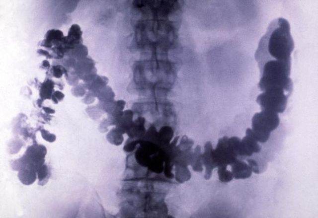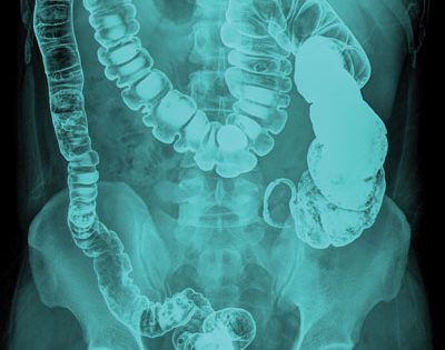
Ionic hyperosmolaric contrast media may no longer be used intravenously due to their high osmolality with related side effects and are thus used primarily in gastrointestinal diagnostic procedures. Iodinated contrast media can be separated into two groups: ionic or non-ionic preparations. Iodine-containing contrast agents include: iobitridol, iodamide, iodixanol, iohexol, iomeprol, iopamidol, iopanoic acid, iopentol, iopodate, iopromide, iotalamic acid, iotrolan, iotroxic acid, ioversol, ioxaglic acid, ioxitalamic acid, lysine amidotrizoate, meglumine amidotrizoate and sodium amidotrizoate, metrizamide and metrizoate.

A small prospective cohort study found no increased risk after diagnostic upper gastrointestinal tract fluoroscopic examination with barium swallow in pregnancy ( Han 2011). Therefore, no damage to the fetus is expected from this contrast medium during pregnancy. This insoluble compound is not absorbed by the digestive tract. Stefanie Hultzsch, in Drugs During Pregnancy and Lactation (Third Edition), 2015 2.20.2 Contrast media Bariumsulfate contrast mediumīarium sulfate is used for radiologic opacification of the stomach and intestinal tract. In the United States, barium is usually assigned a “white” value in Japan, barium often is assigned a “black” value. Depending on the radiologist, large exposures are assigned either “white” or “black” values. The images are displayed on a monitor or flat panel. The relative exposures on the image capture device are digitized and are assigned gray-scale values, from white to black. Today, most radiographs are not obtained using real film as the recording medium, but they use some type of image capture device.

The gastric antrum appears “gray,” not black as a purely air-filled structure would appear. Most of the x-rays, but not all, pass through the antrum, partially exposing the film. Because barium absorbs more x-rays than air, the gastric fundus appears “white.” In this radiograph, the anteriorly located gastric antrum is filled with air and is also “etched in white” by a thin layer of barium. The gastric fundus is posterior to the gastric antrum barium falls into the gastric fundus when the patient lies in a supine position. This image was obtained with the patient recumbent, back against the fluoroscopic tabletop (a supine radiograph). The heavy barium falls to the lowest or most “dependent” portion of the stomach. The relatively exposed areas of the film appear as shades of black the relatively unexposed areas of the film appear as shades of white. X-rays easily pass through GI structures filled with air and expose the film. X-rays passing through barium are blocked and do not reach the film. 15-2, whereas the antrum appears gray?īarium sulfate absorbs and scatters x-rays, but air does not.
#Barium exposure x ray plus
Rubesin MD, in Radiology Secrets Plus (Third Edition), 2011 6 Why does the gastric fundus appear white in Fig.
#Barium exposure x ray software
The number and location of markers depend on the software program being used ( Box 18-6). The soft tissue can be determined as the difference between the template and bone. With the double-scan technique, the denture base resin and artificial teeth can be reconstructed for planning purposes. The software program uses the markers to correlate the images. The patient is scanned wearing the template, and then the template is removed from the patient's mouth and scanned separately. In this technique, reference markers (radiopaque material) are embedded into the radiopaque template. Some planning software programs require the use of a “double-scan” process as the protocol for their software, which exhibits less scatter than the single-scan technique. Poor mixing will result in a nonhomogeneous mixture that exhibits areas of high radiolucency. This allows for differentiation of the teeth from the soft tissue. If a soft tissue (flapless surgery) template is to be made, teeth are ideally identified with a 20% BaSO 4 solution, and the base (soft tissue) uses a 10% mix. Single Scan.īarium sulfate is used to identify the teeth from the diagnostic wax-up in a 20% BaSO 4 solution. The protocol should be obtained before the scan to avoid complications with integration of the CT data into the scanning program ( Box 18-5). The composition and design of the radiopaque template depend on the type of software used during the planning.

Most planning software today uses a radiopaque template with a single-scan technique (discussed later for partial and fully edentulous). Misch, in Dental Implant Prosthetics (Second Edition), 2015 Single-Scan versus Double-Scan Technique


 0 kommentar(er)
